- October 8, 2025
- Posted by: Qamar Un Nisa
- Category: Uncategorized
In today’s fast-evolving world of medical education, technology continues to transform the way students and professionals explore the complexities of the human body. One of the most advanced innovations in this field is the Anatomage Table a state-of-the-art 3D anatomy and physiology learning platform that provides an interactive, lifelike experience. It has become an essential educational tool for medical schools, universities, hospitals, and research centers worldwide.
What is the Anatomage Table?
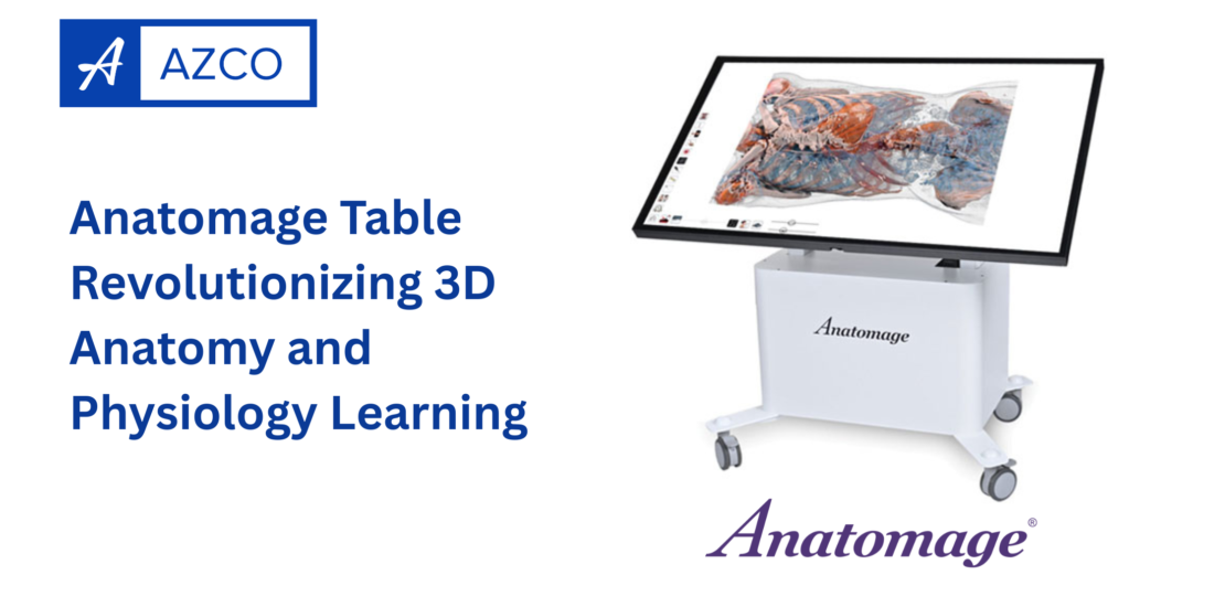
The Anatomage Table is the world’s first and most advanced 3D anatomy visualization and virtual dissection system. It functions like a life-sized digital cadaver, allowing users to explore every aspect of human anatomy in extraordinary detail. Unlike traditional cadavers, which can be limited by availability and preservation issues, the Anatomage Table offers a clean, precise, and endlessly reusable way to study the human body.
This cutting-edge table uses real human data obtained through high-resolution scans, creating anatomically accurate 3D representations of the human body. Users can virtually dissect layer by layer, zoom in on organs, study tissue structures, and even simulate physiological functions all through an intuitive touch-screen interface.
How the Anatomage Table Transforms Anatomy Learning
For decades, medical students have relied on textbooks, plastic models, and cadaver dissections to learn human anatomy. While these methods are valuable, they often have limitations in clarity, accessibility, and interactivity. The Anatomage Table overcomes these challenges by combining advanced visualization technology with realistic human data, making anatomy learning more immersive and efficient.
- Interactive 3D Exploration
The table’s 3D interface allows users to rotate, zoom, and dissect the human body in any direction. Students can visualize complex structures such as muscles, blood vessels, and nerves with complete control. This interactivity fosters a deeper understanding of spatial relationships within the body something that flat textbook images simply cannot achieve. - Real Human Anatomy in High Resolution
The Anatomage Table is built from actual human cadaver scans using advanced imaging techniques like CT and MRI. This means the anatomical structures are not computer-generated but based on real human data, ensuring exceptional accuracy. Students can switch between male and female models or view special pathological cases to see how diseases alter anatomy. - Virtual Dissection Without Limitations
Unlike traditional dissection labs, which require physical cadavers and complex preparation, the Anatomage Table provides an ethical and convenient alternative. Users can make endless virtual dissections, reverse cuts instantly, and highlight specific body systems for focused learning. This repeatable and clean process allows educators to conduct practical sessions without the constraints of real cadaver labs. - Dynamic Physiology Simulations
Beyond anatomy, it also incorporates physiological and clinical features. It includes visualizations of organ functions, blood flow, and interactive case studies that connect anatomical structures with real-life medical scenarios. Students can simulate surgical procedures, study cross-sectional imaging, and correlate anatomy with pathology bridging the gap between classroom learning and clinical practice.
Educational Benefits of the Anatomage Table
The Anatomage Table is more than a technological marvel it is a powerful learning platform designed to improve comprehension, engagement, and retention in medical education.
- Enhanced Visualization and Comprehension
The human body is complex, and understanding its systems requires more than memorization. The Anatomage Table offers an interactive 3D perspective that helps students grasp how organs and systems interconnect. This visual clarity leads to a stronger conceptual understanding. - Active and Collaborative Learning
The large touch-screen interface of the Anatomage Table encourages group learning. Multiple students can gather around the table, discuss structures, and interact simultaneously. This collaborative environment fosters discussion, teamwork, and active engagement. - Bridging Anatomy and Clinical Practice
The Anatomage Table’s integration of radiological imaging allows students to compare 3D anatomy with CT or MRI scans. This feature helps future doctors interpret medical images more effectively and understand clinical correlations earlier in their training. - Accessibility and Sustainability
Traditional cadaver labs require special facilities, preservation chemicals, and ethical considerations. In contrast, the Anatomage Table eliminates these constraints, providing a reusable, maintenance-free, and environmentally friendly solution.
Features that Make the Anatomage Table Stand Out
The Anatomage Table offers a wide range of features that make it the gold standard for 3D anatomy education:
- Full-Body Anatomical Models
Each table comes with highly detailed male and female anatomical datasets that allow for complete body dissections from head to toe. - Regional and System-Based Views
Users can isolate specific body systems — skeletal, muscular, cardiovascular, nervous, and more — or study regional anatomy such as the head, thorax, abdomen, or limbs. - Pathological Case Library
The platform includes real patient scans with clinical pathologies, giving learners exposure to real-world conditions and diagnostic challenges. - Integration with Medical Imaging
Educators and students can upload their own CT and MRI scans to analyze patient cases directly on the Anatomage Table. - Annotation and Quiz Tools
Teachers can create customized quizzes, label structures, and conduct assessments, turning the table into an interactive teaching and testing tool.
Applications in Education and Healthcare
The Anatomage Table has found its place not only in medical schools but also in nursing colleges, physiotherapy departments, dental institutions, and hospitals. Here are some of its practical applications:
- Medical and Allied Health Education:
Used for teaching anatomy, physiology, radiology, and surgical training. - Surgical Planning and Demonstration:
Surgeons can use the Anatomage Table to visualize patient anatomy and plan complex procedures before operating. - Patient Education:
Doctors can use the table to explain diagnoses and treatment plans to patients using clear 3D visuals. - Research and Development:
Researchers can study anatomical variations and pathological changes in detail using the high-resolution datasets.
Why Educational Institutions Choose the Anatomage Table
Many prestigious medical universities and teaching hospitals around the world have adopted the Anatomage Table as part of their curriculum. The reasons are clear: it enhances teaching quality, reduces costs associated with cadaver management, and keeps students engaged with interactive learning.
Furthermore, the Anatomage Table aligns with modern digital education trends, making it ideal for blended and virtual learning environments. It supports remote teaching by allowing screen sharing and online demonstrations, ensuring continuity in education even when physical access to labs is limited.
The Future of Anatomy Learning
As the field of medical education continues to embrace digital transformation, this represents the future of anatomical science. Its blend of realism, interactivity, and accessibility makes it an indispensable tool for educators and students alike.
By combining the precision of real human anatomy with the flexibility of virtual learning, it’s not only enhances understanding but also inspires curiosity and innovation. It is a testament to how technology can make learning more engaging, sustainable, and effective.
Conclusion
The Anatomage Table is more than just a piece of technology it is a gateway to a new era of medical education. With its 3D anatomy visualization, virtual dissection capabilities, and physiological simulations, it empowers students to explore the human body like never before. Whether used in classrooms, hospitals, or research labs, the Anatomage Table stands as a powerful learning platform that bridges the gap between theory and clinical practice.
As education continues to evolve, tools like this will play a crucial role in shaping the next generation of healthcare professionals equipped with deeper understanding, sharper skills, and a passion for discovery.