

- August 17, 2026
- Posted by: Qamar Un Nisa
- Category: Uncategorized
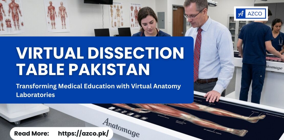
Medical education is rapidly moving toward more interactive and technology-driven learning. As medical universities, teaching hospitals, and healthcare institutions in Pakistan look for innovative ways to improve anatomy education, the virtual dissection table Pakistan market is gaining attention.
Traditional anatomy teaching remains an essential part of medical education, but digital technologies now provide students with new ways to explore the human body. A virtual dissection table allows learners to interact with three-dimensional anatomy, examine anatomical structures from multiple angles, perform virtual dissections, and connect anatomy with clinical imaging.
For institutions planning to establish a modern virtual anatomy laboratory or digital anatomy laboratory, a virtual dissection table can become an important educational technology investment.
What Is a Virtual Dissection Table?
A virtual dissection table is an interactive digital platform designed to help students and educators study human anatomy in three dimensions. Instead of relying exclusively on physical specimens, students can explore digital representations of human anatomy on a large interactive touchscreen.
The technology allows users to rotate, zoom, isolate, dissect, and examine different anatomical structures. Depending on the platform, users can also explore muscles, bones, organs, blood vessels, nerves, and other structures individually or as part of the complete human body.
This approach can make anatomy lessons more visual, interactive, and accessible while giving instructors additional tools for demonstrations and practical teaching.
Virtual Dissection Table Pakistan: Why Is It Becoming Important?
Medical colleges and universities in Pakistan are increasingly interested in modern teaching technologies that can support conventional anatomy education.
A virtual dissection table Pakistan solution can provide several advantages for institutions:
- Interactive 3D anatomy visualization
- Virtual dissection without physical specimen preparation
- Easy identification of anatomical structures
- Repeated demonstrations and practical sessions
- Group-based classroom learning
- Integration of anatomy with medical imaging
- Flexible revision for students
- Modern technology for anatomy laboratories
Rather than replacing every traditional teaching method, virtual anatomy technology can complement lectures, textbooks, anatomical models, and cadaver-based learning.
Building a Modern Virtual Anatomy Laboratory
A virtual anatomy laboratory is an educational environment where students use digital technologies to study anatomy through interactive visualization and simulation.
A virtual anatomy laboratory can include:
- 3D anatomy visualization systems
- Virtual dissection tables
- Interactive anatomical models
- Digital imaging resources
- CT and MRI case libraries
- Anatomy teaching software
- Large-format interactive displays
- Digital learning resources
The objective is to create an environment where students can actively explore anatomy rather than simply observing static diagrams.
For medical universities, this can make practical anatomy sessions more engaging and provide instructors with new ways to explain complicated anatomical relationships.
Digital Anatomy Laboratory: The Future of Anatomy Education
A digital anatomy laboratory uses technology to create a modern learning environment for anatomy and related healthcare subjects.
Instead of depending entirely on physical specimens and printed diagrams, students can access interactive digital anatomy and examine structures repeatedly.
For example, an instructor can demonstrate the cardiovascular system to an entire class by displaying the heart and major blood vessels in 3D. Students can observe the structures from different angles and explore their anatomical relationships.
Similarly, the nervous system, musculoskeletal system, respiratory system, digestive system, and other body systems can be studied using interactive visualization.
This approach can be particularly useful when teaching structures that are difficult to understand through two-dimensional images.
Key Features of a Virtual Dissection Table
1. Interactive 3D Human Anatomy
The ability to visualize anatomy in three dimensions is one of the most important advantages of a virtual dissection system.
Students can rotate the digital body, zoom into specific regions, and isolate anatomical structures for detailed examination.
2. Virtual Dissection Table Pakistan
Students can perform digital dissections to understand the relationship between superficial and deeper structures.
Unlike a physical specimen, a digital model can be restored and explored repeatedly, allowing students to revisit the same anatomical region during revision.
3. Layer-by-Layer Exploration
A digital anatomy platform can allow students to move through anatomical layers and examine structures progressively.
This can help demonstrate relationships between skin, muscles, bones, vessels, nerves, and organs.
4. Clinical Anatomy and Medical Imaging
Modern anatomy platforms can connect anatomical education with clinical imaging. CT and MRI datasets can help students understand how anatomy appears in diagnostic imaging.
This creates a bridge between basic anatomy and clinical learning.
5. Interactive Classroom Teaching
A large touchscreen virtual dissection table allows instructors to demonstrate anatomy to groups of students.
Teachers can highlight structures, rotate models, explain anatomical relationships, and guide students through virtual dissections during practical sessions.
Benefits of a Virtual Anatomy Laboratory for Medical Colleges
A virtual anatomy laboratory can provide educational benefits for both students and instructors.
Better Visualization
Three-dimensional visualization can help students understand spatial relationships that may be difficult to interpret from traditional two-dimensional diagrams.
Repeated Practice
Students can repeatedly explore anatomical structures without consuming physical specimens.
Improved Engagement
Interactive technology encourages students to participate actively during anatomy sessions.
Flexible Learning
Digital anatomy can be used for classroom demonstrations, practical sessions, group activities, presentations, and individual revision.
Integration With Clinical Education
Digital anatomy and medical imaging can help students understand the connection between anatomical knowledge and clinical practice.
Virtual Dissection Table vs Traditional Anatomy Methods
A virtual dissection table should not necessarily be considered a complete replacement for traditional anatomy education.
Cadaveric dissection provides important hands-on experience and remains valuable in medical education. However, digital anatomy can complement this experience by providing an additional method for visualization and repeated exploration.
For example, students may use cadaveric specimens for hands-on learning and then use a virtual dissection table to revisit the same region in 3D.
This combination can provide students with both physical and digital learning experiences.
Who Can Benefit From a Digital Anatomy Laboratory?
A digital anatomy laboratory can be useful for a wide range of educational institutions and healthcare programs.
Virtual Dissection Table Pakistan in Medical Universities
Medical universities can use digital anatomy technology to enhance anatomy lectures, practical sessions, demonstrations, and student learning.
Virtual Dissection Table Pakistan in Medical Colleges
Medical colleges can establish dedicated virtual anatomy laboratories to modernize anatomy teaching.
Teaching Hospitals
Teaching hospitals can use interactive anatomy and imaging resources for medical education and training.
Dental Institutions
Dental students can use 3D visualization to study head and neck anatomy and related structures.
Allied Health Programs
Programs such as physiotherapy, radiology, nursing, and other health sciences can benefit from interactive anatomy resources according to their curriculum.
Choosing a Virtual Dissection Table in Pakistan
When evaluating a virtual dissection table Pakistan solution, institutions should consider more than the hardware.
Important factors include:
- 3D anatomy content – Check the quality and depth of anatomical datasets.
- Software capabilities – Evaluate dissection, visualization, imaging, and teaching features.
- Display and hardware – Consider screen size, resolution, durability, and usability.
- Training – Ensure faculty members receive proper training.
- Installation – Confirm that installation and commissioning support are available.
- Technical support – Consider warranty, maintenance, updates, and after-sales service.
- Curriculum integration – Evaluate how the system will fit into existing anatomy courses.
Why Invest in a Virtual Anatomy Laboratory?
The goal of a virtual anatomy laboratory is not simply to introduce new technology. It is to improve the way students interact with anatomical information.
Medical students need to understand not only what an anatomical structure looks like but also where it is located, how it relates to surrounding structures, and how anatomy connects to clinical applications.
Interactive digital anatomy can help create a learning environment where students can explore these relationships visually and repeatedly.
For institutions in Pakistan, establishing a digital anatomy laboratory can also demonstrate a commitment to modern medical education and technology-enabled learning.
Conclusion
The demand for innovative medical education technology is creating new opportunities for virtual dissection table Pakistan solutions. By combining interactive 3D anatomy, virtual dissection, digital visualization, and clinical imaging, these systems can provide medical institutions with a powerful addition to conventional anatomy teaching.
A well-designed virtual anatomy laboratory can help students visualize complex structures, practice repeatedly, and develop a stronger understanding of three-dimensional anatomy. Similarly, a digital anatomy laboratory can connect traditional anatomy education with modern digital learning methods.
For medical universities, colleges, teaching hospitals, and healthcare institutions considering an upgrade to their anatomy teaching facilities, investing in a virtual dissection system can be an important step toward a more interactive and technology-enabled educational environment.
Looking to establish a virtual anatomy laboratory in Pakistan? Contact AZCO to explore advanced 3D anatomy and virtual dissection solutions designed for modern medical education.